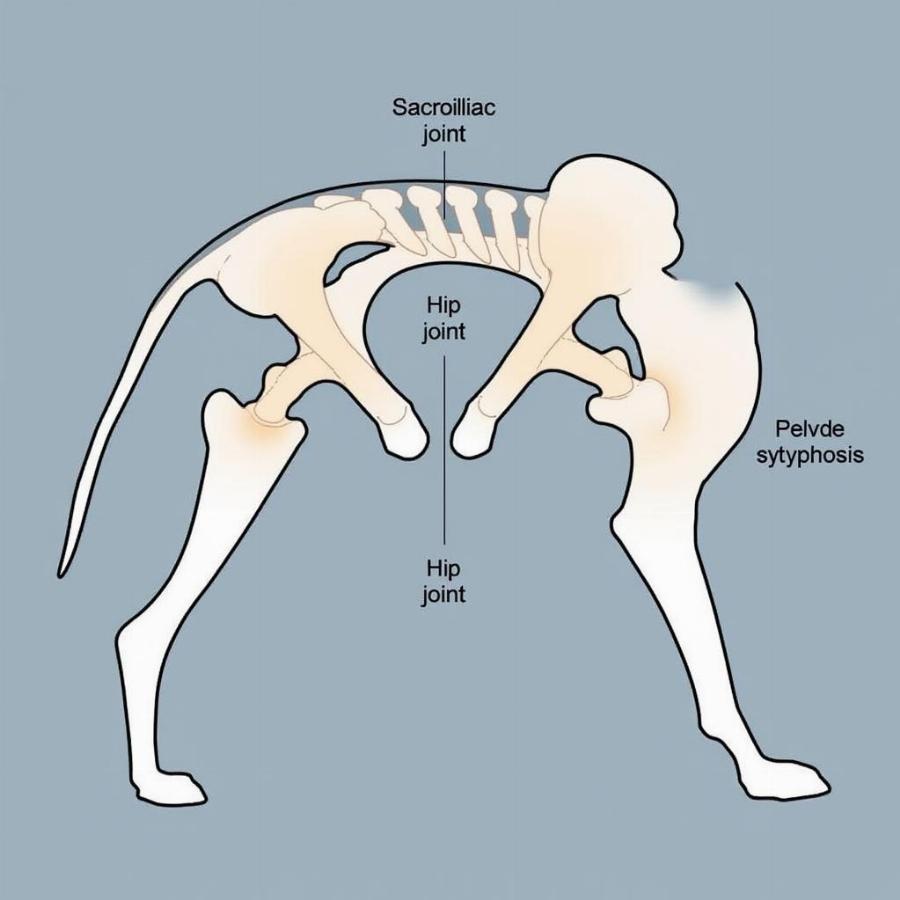
Dog pelvis anatomy is a crucial aspect of canine health and well-being. Understanding the structure and function of this complex bone structure can help owners recognize potential issues, understand diagnostic procedures, and appreciate the biomechanics of their dog’s movement. This article will delve into the intricacies of the dog pelvis, providing valuable insights for both pet owners and enthusiasts.
Decoding the Dog Pelvis: Bones, Joints, and Functions
The dog pelvis, also known as the pelvic girdle, is a complex structure composed of three paired bones: the ilium, ischium, and pubis. These bones fuse together to form the hip joint, a ball-and-socket joint where the head of the femur (thigh bone) articulates with the acetabulum of the pelvis. The pelvis plays several vital roles, including supporting the hind limbs, protecting the abdominal organs, and providing attachment points for muscles involved in locomotion, urination, and defecation.
The Ilium: The Wing-Like Bone
The ilium is the largest of the three pelvic bones and forms the cranial (front) part of the pelvis. It has a wing-like projection called the iliac crest, which can be easily felt on either side of the dog’s lower back. The ilium provides attachment points for several important muscles, including those that flex the hip and extend the back.
The Ischium: Supporting the Sit Bone
The ischium forms the caudal (rear) part of the pelvis and provides the bony prominence known as the ischial tuberosity – the “sit bone”. This bone is crucial for supporting the dog’s weight when sitting and also serves as an attachment point for muscles involved in extending the hip and flexing the knee.
The Pubis: Completing the Pelvic Ring
The pubis is the smallest of the three pelvic bones and forms the ventral (bottom) part of the pelvis. The two pubic bones join together at the pelvic symphysis, a cartilaginous joint that allows for slight movement. This flexibility is particularly important during pregnancy and parturition.
 Dog Pelvis Joints
Common Issues Related to Dog Pelvis Anatomy
Several health problems can affect the dog pelvis, including hip dysplasia, pelvic fractures, and osteoarthritis. Hip dysplasia, a common developmental disorder, affects the hip joint and can lead to pain, lameness, and decreased mobility. Pelvic fractures, often caused by trauma, can be serious and require surgical intervention. Osteoarthritis, a degenerative joint disease, can affect the hip joint and cause pain and stiffness.
Diagnosing Pelvic Problems in Dogs
Veterinarians use various diagnostic tools to evaluate the dog pelvis, including physical examination, radiographs (X-rays), computed tomography (CT scans), and magnetic resonance imaging (MRI). These tools allow veterinarians to visualize the bones and joints of the pelvis, identify abnormalities, and make an accurate diagnosis.
How a Dog’s Pelvis Affects its Gait
The structure and function of the dog pelvis are intricately linked to its gait and movement. The angle of the pelvis, the length of the bones, and the strength of the surrounding muscles all contribute to the dog’s ability to walk, run, and jump. anatomy of a dog’s rear leg provides further insights into the mechanics of the hind limbs.
Conclusion
Understanding dog pelvis anatomy is essential for responsible pet ownership. By recognizing the importance of this complex structure, owners can better understand their dog’s health, movement, and potential vulnerabilities. This knowledge empowers owners to make informed decisions about their dog’s care, ensuring a happy, healthy, and active life. Knowing how many bones a dog has in total (how much bones does a dog have) can also be helpful in understanding overall skeletal health.
FAQ
- What are the main bones of the dog pelvis? The ilium, ischium, and pubis.
- What is the function of the acetabulum? It forms the socket part of the hip joint.
- What is hip dysplasia? A common developmental disorder affecting the hip joint.
- How are pelvic problems diagnosed in dogs? Through physical exams, X-rays, CT scans, and MRI.
- How does the pelvis affect a dog’s gait? It influences the dog’s ability to walk, run, and jump.
- What is the pelvic symphysis? The cartilaginous joint connecting the two pubic bones.
- Where is the ischial tuberosity located? At the caudal part of the pelvis, forming the “sit bone”.
Beaut Dogs is your trusted source for comprehensive and reliable information on the world of canine companions. We provide expert guidance on all aspects of dog ownership, from breed selection to specialized care. vertebral anatomy dog offers a deeper understanding of the canine skeletal system. When you need expert advice, don’t hesitate to reach out to Beaut Dogs at [email protected] for detailed and accurate answers. anatomy female dog provides specific information related to the female dog’s anatomy.